| 下腰痛患者终板Modic改变在腰椎上的分布特点 |
腰椎终板改变即Modic改变,是指MRI上可见的邻近椎体终板退变引起的骨髓信号改变。Modic等[1]于1988年将其分为Ⅰ型、Ⅱ型及Ⅲ型3种类型,之后根据同一终板受累的不同类型及不同比例,又增加了7种混合型,如Ⅰ-Ⅱ型、Ⅱ-Ⅲ型等[2]。下腰痛是临床常见症状,与许多因素相关,包括Modic改变及腰椎间盘退变,尤其是Modic改变Ⅰ型,但仍有争议。许多研究[3-8]侧重于分析下腰痛与Modic改变类型或椎间盘退变的关系,对下腰痛直接的生物学机制,包括力学作用及进一步发生的终板血供改变研究相对较少。
终板作为椎体与移植物的交界面,在压力性负荷分布方面起重要作用。薄弱的终板可增加椎间融合器沉陷的风险。了解终板完整性的改变可减少移植物沉陷的风险,改善椎间融合效果[9]。本研究探讨腰痛患者腰椎终板Modic改变的分布形式,并根据Modic改变在终板的分布特点阐述力学作用。
1 资料与方法 1.1 一般资料选择2016年10—12月间在我院接受腰椎MRI检查且合并Modic改变的118例(1 180个终板)下腰痛患者作为研究对象,男56例,女62例;年龄20~85岁,平均52.69岁。纳入标准:①年龄20~90岁,合并下腰痛;②已接受腰椎MRI检查;③腰椎终板存在Modic改变。排除标准:①年龄 < 20岁或>90岁;②既往有腰椎手术史;③合并腰椎骨折、肿瘤、感染;④合并腰椎严重的先天性异常、完全或不完全的移行椎、严重的脊柱侧弯等。
1.2 仪器与方法使用GE Signa HDxt 1.5 T MRI扫描仪。患者取仰卧位,腰椎检查采用全脊柱阵列线圈(HD 8Ch CTL)。扫描序列及参数:FSE T1WI TR/TE 450 ms/9.4 ms;FSE T2WI TR/TE 2 560 ms/120 ms;T2压脂序列FSFSE T2WI TR/TE 3 000 ms/102 ms;层厚4 mm,层距0.5 mm,矩阵320×192(T1WI及T2WI)、384×192(T2压脂序列),FOV 320 mm×320 mm。
1.3 图像分析 1.3.1 Modic改变分型采用Modic等制订的原始标准,即依据MRI上终板及终板下骨质的信号改变对Modic改变分型:Ⅰ型,T1WI呈低信号,T2WI呈高信号;Ⅱ型,T1WI、T2WI均呈高信号;Ⅲ型,T1WI、T2WI均呈低信号。混合型Modic改变也列入记录范围内。
1.3.2 终板位置包括腰椎间盘上下的10个终板,即从L1椎体下终板至S1椎体上终板。记录合并Modic改变终板所在的腰椎水平、相对于邻近椎间盘的位置,以及Modic改变的类型、部位和程度。
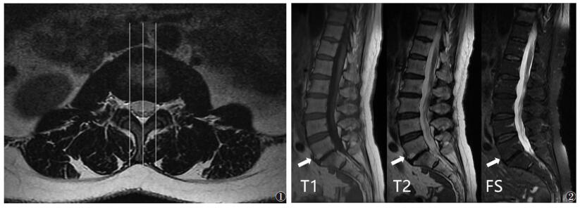
1.3.3 终板分部不同椎间盘的大小、纤维环与髓核比例不尽相同,而硬膜囊左右侧缘对应终板的位置相似。本研究每例患者取13幅图像,几乎能代表椎体全部横径。硬膜囊左右两侧距正中3~4个层面。终板分为硬膜囊左右两侧及正中矢状位3个层面;将椎体终板前后缘3等分,即前部、中部和后部。观察内容:①终板3个矢状位层面是否合并Modic改变及其类型;②Modic改变在每个层面中的发生部位;③Modic改变的程度,无、局限(< 25%椎体高度)、广泛(≥25%椎体高度),以层面最大程度为标准(图 1,2)。
 |
| 图 1 T2WI轴位。从左向右3条矢状位直线分别代表硬膜囊右侧、正中及硬膜囊左侧3个矢状位层面 图 2 女,51岁,硬膜囊右侧矢状位层面。Modic改变Ⅱ型,L5~S1上下终板前部见T1WI高信号、T2WI高信号影,压脂序列未见异常信号;上终板Modic改变程度为广泛(箭头),下终板为局部 |
1.4 统计学分析
采用SPSS 24.0统计学软件进行处理。Modic改变在腰椎终板的分布行χ2检验。以P < 0.05为差异有统计学意义。
2 结果118例1 180个终板中,514个发生Modic改变,发生率43.56%。
2.1 Modic改变在腰椎的分布从L1~2、L2~3、L3~4、L4~5、L5~S1,Modic改变个数分别为35个(6.81%)、47个(9.14%)、88个(17.12%)、145个(28.21%)和199个(38.72%)。各节段间Modic改变的发生率差异均有统计学意义(均P < 0.01)。合并Modic改变的终板中,344个(66.93%)终板位于下腰段(L4~5和L5~S1),明显高于上腰段(33.07%,170/514)。
2.2 Modic改变类型Modic改变Ⅱ型110例(93.22%),Ⅰ型39例(33.45%),Ⅲ型4例(3.39%)。7种混合型Modic改变作为一个整体统计,共48例(40.68%)可见。514个终板中Ⅰ型73个(14.20%),Ⅱ型341个(66.34%),Ⅲ型4个(0.78%),混合型96个(18.68%)。
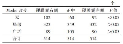
2.3 Modic改变在同一终板的分布 2.3.1 同一终板矢状位层面分布(表 1)| 表 1 矢状位层面不同程度Modic改变的数量 |
 |
514个终板中454个(88.33%)终板正中受累,与硬膜囊左、右侧Modic改变的分布差异均有统计学意义(均P < 0.05),而硬膜囊左、右侧Modic改变数量差异无统计学意义(P>0.05)。局限与广泛Modic改变的数量差异无统计学意义(P>0.05)。
2.3.2 同一终板前、中、后部分布514个终板中464个(90.27%)前部受累,175个(34.05%)中部受累,282个(54.86%)后部受累,三者差异均有统计学意义(均P < 0.05),且两两之间差异均有统计学意义(均P < 0.05)。终板前部Modic改变数量最多,其次为后部,中部受累较少。
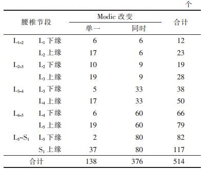
2.4 Modic改变在同一椎间盘上下终板的分布(表 2)| 表 2 Modic改变在同一椎间盘上下终板的分布 |
 |
514个终板中发生在椎间盘上终板217个,下终板297个。同一椎间盘上下终板同时发生Modic改变376个,远高于仅累及单一上下终板数(138个)。Modic改变在L5~S1水平下终板显著多于上终板(P < 0.05)。
3 讨论Modic改变为椎体终板在MRI上信号的改变,Ⅱ型被认为是骨髓的脂肪取代,Ⅰ型则为炎症反应阶段[10-11],两者均与临床下腰痛有关。Ⅰ型是引起下腰痛的主要原因,但仍有争议[10]。3种Modic改变可相互转换,尤其是Ⅰ型与Ⅱ型的关系更为密切[2, 7]。Modic改变与机械负荷[10, 12]和椎间盘退变[1, 5-6]相关,终板营养通路破坏及髓核压力降低被认为是椎间盘退变的2个关键机制[12]。正常终板的中央椎体小梁间隙较宽,其内血管伸入骨质内,终板也相对较薄[13-15]。中央薄弱的终板易受损,血供及营养受阻,进而导致椎间盘退变。Dolan等[16]发现受损的终板突入椎体使得髓核内水分减少,压力降低,负荷逐渐转移至纤维环,使其所受压力增大。增大的终板和软骨下骨小梁压力进一步加重微骨折,导致更严重的终板损伤。推测这种恶性循环更易先起于薄弱的中央终板,而髓核内的压力对细微的容量变化很敏感,损伤越重,髓核压力越低,后期纤维环承受压力越大[16]。Liu等[9]发现,终板的弹性和承受力由中央向外周增加,作用于邻近椎间盘的压力也在中央髓核处最小,在外周纤维环最大,而当终板和椎间盘退变时,弹性和承受力均下降,外周甚于中央。原先外周纤维环可承受较大负荷的能力较中央髓核更明显下降,此时压力性负荷重新分配,终板整体损伤加重,尤其是中央终板。不同矢状位层面对Modic改变的程度并无影响,推测力学的改变可能并非唯一的影响因素,有待进一步研究。本研究发现,Modic改变更多发生在终板前部,其次为后部,中部累及较少。Hulme等[13]发现,前方和中央区域的终板最薄,后方最厚,且前方及中央区的小梁密度较低。终板前后部由于长期、反复的伸展和屈曲动作,承受负荷的机会更多,相较于中央更易受累。腰椎的伸展及骨折可使负荷转移至前部椎体皮质[16],这也是老年人压缩性骨折更常见的部位。可以推断,退变发生后,终板中央区域由于髓核内压力降低致损伤加重更易受累,而其前后部,尤其是前部,因长期、反复的伸展和屈曲运动使承受的负荷较其他部位增长更显著。
椎体上终板较下终板更薄,且下终板随腰椎水平的降低而增厚[14]。邻近椎间盘上方终板的骨小梁密度比下方高,当邻近椎间盘压迫椎体时,下终板常较上终板先受损。终板在下腰段弹性和承受力增加,在椎间盘下终板亦如此[9, 15]。本研究发现,Modic改变好发L5~S1水平,受累下终板数明显较上终板数多,而同时累及上下终板数较单一终板多。腰椎水平可能通过纤维环高度及髓核容量的降低产生影响[16],在退行性椎间盘疾病中下腰段承受负荷最大[15, 17],这可能是Modic改变好发于下腰段的原因。
总之,Modic改变好发于下腰段,常同时累及邻近椎间盘上下终板。Modic改变以Ⅱ型为主。同一终板的不同区域对Modic改变程度无明显影响,力学的改变可能并非唯一的影响因素。
| [1] |
Modic MT, Masaryk TJ, Ross JS, et al. Imaging of degenerative disk disease[J]. Radiology, 1988, 168: 177-186. DOI:10.1148/radiology.168.1.3289089 |
| [2] |
Xu L, Chu B, Feng Y, et al. Modic changes in lumbar spine:prevalence and distribution patterns of end plate oedema and end plate sclerosis[J]. Br J Radiol, 2016, 89: 20150650. DOI:10.1259/bjr.20150650 |
| [3] |
Feng Z, Liu Y, Wei W, et al. Type Ⅱ modic changes may not always represent fat degeneration:A study using MR fat suppression sequence[J]. Spine, 2016, 41: 987-994. DOI:10.1097/BRS.0000000000001395 |
| [4] |
Wang Y, Videman T, Battié MC. Modic changes:prevalence, distribution patterns and association with age in Caucasian men[J]. Spine J, 2012, 12: 411-416. DOI:10.1016/j.spinee.2012.03.026 |
| [5] |
Albert HB, Briggs AM, Kent P, et al. The prevalence of MRI-defined spinal pathoanatomies and their association with modic changes in individuals seeking care for low back pain[J]. Eur Spine J, 2011, 20: 1355-1362. DOI:10.1007/s00586-011-1794-6 |
| [6] |
Yu LP, Qian WW, Yin GY, et al. MRI assessment of lumbar intervetebral disc degeneration with lumbar degenerative disease using the pfirrmann grading systems[J]. PLoS One, 2012, 7: 48074. DOI:10.1371/journal.pone.0048074 |
| [7] |
Hutton MJ, Bayer JH, Powell JM. Modic vertebral body changes:the natural history as assessed by consecutive magnetic resonance imaging[J]. Spine, 2011, 36: 2304-2307. DOI:10.1097/BRS.0b013e31821604b6 |
| [8] |
Muftuler LT, Jarman JP, Yu HJ, et al. Association between intervertebral disc degeneration and endplate perfusion studied by DCE-MRI[J]. Eur Spine J, 2015, 24: 679-685. DOI:10.1007/s00586-014-3690-3 |
| [9] |
Liu J, Hao L, Suyou L, et al. Biomechanical properties of lumbar endplates and their correlation with MRI findings of lumbar degeneration[J]. J Biomech, 2016, 49: 586-593. DOI:10.1016/j.jbiomech.2016.01.019 |
| [10] |
Modic MT. Modic type 1 and 2 changes[J]. J Neurosurg Spi ne, 2007, 6: 150-151. |
| [11] |
Kuisma M, Karppinen J, Niinimaki J, et al. Modic changes in endplates of lumbar vertebral bodies:prevalence and association with low back and sciatic pain among middle-aged male work ers[J]. Spine, 2007, 32: 1116-1122. DOI:10.1097/01.brs.0000261561.12944.ff |
| [12] |
Holm S, Holm AK, Ekstrom L, et al. Experimental disc degene ration due to endplate injury[J]. J Spinal Disord Tech, 2004, 17: 64-71. DOI:10.1097/00024720-200402000-00012 |
| [13] |
Hulme PA, Boyd SK, Ferguson SJ. Regional variation in verteb ral bone morphology and its contribution to vertebral fracture strength[J]. Bone, 2007, 41: 946-957. DOI:10.1016/j.bone.2007.08.019 |
| [14] |
Zhao FD, Pollintine P, Hole BD, et al. Vertebral fractures usua lly affect the cranial endplate because it is thinner and suppo rted by less-dense trabecular bone[J]. Bone, 2009, 44: 372-379. DOI:10.1016/j.bone.2008.10.048 |
| [15] |
Hou Y, Luo Z. A study on the structural properties of the lu mbar endplate:histological structure, the effect of bone density, and spine level[J]. Spine, 2009, 34: 427-433. DOI:10.1097/BRS.0b013e3181a2ea0a |
| [16] |
Dolan P, Luo J, Pollintine P, et al. Intervertebral disc decompr ession following endplate damage:implications for disc degener ation depend on spinal level and age[J]. Spine, 2013, 38: 14731481. |
| [17] |
West W, West KP, Younger EN, et al. Degenerative disc disease of the lumbar spine on MRI[J]. West Indian Med J, 2010, 59: 192-195. |
 2018, Vol. 16


