 2021, Vol. 51


GATA家族是经典的转录因子家族,结构上具有保守的Ⅳ型锌指结构域,因特异性的与靶基因启动子的核苷酸序列T/A(GATA)A/G结合而得名[1-2]。1989年,Tsai等[3]首次在鸡中克隆获得GATA1基因,随后GATA家族在各物种中展开研究。在脊椎动物中一般具有6个成员(GATA1-6),根据它们的系统进化和组织表达特征,GATA家族分为GATA1/2/3和GATA4/5/6两个亚家族[4]。GATA1/2/3亚家族主要表达于外胚层,是造血系统和中枢神经系统分化和发育所必需的;GATA4/5/6亚家族在中内胚层的器官中表达,如心脏、肝脏、肺、生殖腺等,它们对组织特异性基因的表达起关键的调控作用[5-7]。
在无脊椎动物中,虽然GATA家族成员的数目不同,如在果蝇(Drosophila melanogaster)中有5个,在秀丽隐杆线虫(Caenorhabditis elegans)中有11个,在其它物种中一般小于5个[8],但根据系统进化分析它们都分属于GATA1/2/3和GATA4/5/6亚家族,且在发育过程中与胚层相关器官的分化和功能维持相关[8-12]。近几年,还发现该家族成员在其它方面的功能,如参与先天免疫,在秀丽隐杆线虫受病原体(如铜绿假单胞菌Pseudomonas aeruginosa、鼠伤寒沙门氏菌Salmonella typhimurium、新型隐球菌Cryptococcus neoformans)侵染的过程中,敲除或降低elt-2的表达会使线虫的抗性降低[13];软体动物栉孔扇贝(Chlamys farreri)的GATA和长牡蛎(Crassostrea gigas)的GATA2/3,在成体免疫器官中高表达,且敲降后它们血细胞总数和更新率都显著下降[14-15]。此外,在秀丽隐杆线虫中报道GATA家族的多个成员参与环境应激过程。ELT-2在缺氧、高膳食锌和纳米聚苯乙烯暴露等环境中,通过上调自身表达来调控下游基因的转录以应对环境变化[16-18];ELT-3通过调节角质层胶原相关基因的表达来应对饮食、密度等环境的变化[19];在氧化应激中,ELT-3上调解毒基因谷胱甘肽硫转移酶4(glutathione S-transferase 4, gst-4)的表达[20]。以上研究表明,GATA家族除了在发育中的保守作用外,还有许多功能待探究,包括对环境的应激反应。
单环刺螠(Urechis unicinctus)是具有代表性的螠虫动物(Echiura),隶属于环节动物门(Annelida),是一种海洋无脊椎经济物种,广泛分布于中国、俄罗斯、日本和韩国沿海的潮间带和潮下带的U型洞穴里[19]。螠虫动物生活环境中的硫化物浓度随着含氧量的减少而升高,退潮时甚至高达66 μmol/L[21]。螠虫动物具有较强的硫化物耐受、代谢和利用能力,越来越多的研究者将螠虫动物作为硫化物代谢研究的模式生物[22-24]。前期,李学玉等[25]利用酵母单杂交筛选到GATA B2可能与单环刺螠硫代谢关键基因硫醌氯化还原酶(Sulfide quinone oxidoreductase, sqr)的启动子结合,为了进一步验证其功能,本研究扩增获得单环刺螠GATA B2(UuGATA B2)的cDNA序列,并制备多克隆抗体检测其在成体不同组织和后肠硫化物应激下的表达特征,为深入探讨单环刺螠GATA B2在硫代谢过程中的作用提供基础数据。
1 材料和方法 1.1 实验动物本实验使用的单环刺螠购自青岛市四方路水产品市场,产地为烟台市莱州沿海。将单环刺螠在pH=7.9,盐度32,水温17~19 ℃的海水中通气暂养3 d,期间每天投喂适量小球藻,实验前24 h停止投喂。挑选体表无伤痕、活力旺盛的个体(体重=(30.2±9.8) g)进行实验。
1.2 实验方法 1.2.1 单环刺螠硫化物处理及取样(1) 正常组织取样:挑选上述单环刺螠个体5尾,每尾取体壁、中肠、后肠、肛门囊和体腔液组织,用1×PBS快速清洗后立即放入液氮中冻存,并及时转移至-80 ℃冰箱中保存,用于后续总蛋白提取。
(2) 硫化物处理及取样:挑选60尾上述单环刺螠个体,随机放入6个盛有30 L过滤海水的玻璃容器中。硫化物的添加和浓度测量的方法参照Liu[26]。实验过程中分别在0、0.25、0.5、1、1.5、2、6、12、24、48 h时,从每个容器中随机取一尾单环刺螠解剖并取其后肠组织,样品处理和保存方法同(1)。
1.2.2 总RNA提取与cDNA模板合成取100 mg正常的后肠组织采用酚氯仿抽提法进行总RNA提取,然后根据PrimeScriptTM RT reagent Kit with gDNA Eraser试剂盒(TaKaRa,大连)的操作指南制备cDNA模板。
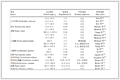
1.2.3 UuGATA B2 cDNA的获得从单环刺螠硫应激的转录组(www.ncbi.nlm.nih.gov/Traces/sra/sra.cgi, accession number: SRA122323)中查找GATA B2 cDNA序列,并在NCBI中的Open Reading Frame Finder工具进行ORF预测,初步确认其ORF序列。然后设计引物GATA B2-F/R(见表 1)用于cDNA的扩增,并送往北京擎科新业公司进行合成。然后以单环刺螠后肠cDNA为模板使用上述合成的引物进行PCR扩增,经过凝胶电泳、胶回收等步骤获得目的片段。再按照pClone007平末端载体(擎科,北京)的操作步骤将回收的目的片段连接到载体上,经转化、培养后挑取单克隆送往生工测序。
|
表 1 本文中所用引物序列 Table 1 The sequences of primers used in this study |
使用ExPASy数据库对目的蛋白的基本理化性质进行分析;使用DNAMAN 6.0软件进行氨基酸同源性比对;利用MEGA 6.0软件以邻接法(Neighbor-joining method,NJ法)构建进化树,Bootstrap设置为1 000;使用SignalP(http://www.cbs.dtu.dk/services/SignalP)预测蛋白质的信号肽;使用TMHMM(http://www.cbs.dtu.dk/services/TMHMM/)预测蛋白质的跨膜结构;使用cNLS Mapper(http://nls-mapper.iab.keio.ac.jp/cgi-bin/NLS_Mapper_form.cgi)预测核定位信号;使用BankiIt将得到的单环刺螠GATA B2相关序列提交GenBank数据库。
1.2.5 UuGATA B2的抗体制备(1) 原核表达载体的构建:设计引物GATA B2-FP/RP(见表 1)扩增GATA B2的ORF,并根据定向表达载体pDE1 Directional Expression Kit(擎科,北京)的使用说明将其连接到pDE1载体上,然后转入DH5α细胞中进行复苏、培养和挑取单克隆,测序验证ORF序列无碱基突变和缺失,完成pDE1-UuGATA B2原核表达载体的构建。
(2) 重组蛋白的原核表达:提取pDE1-UuGATA B2的重组质粒转化入大肠杆菌BL21(DE3)感受态细胞,构建重组表达菌株BL21/pDE1-UuGATA B2。取上述构建好的重组表达菌液100 μL,加入1 mL含有卡那霉素的LB培养基,37 ℃ 300 r/min摇床上复苏过夜。然后以1:100体积比接种于含卡那霉素的100 mL LB培养基中,同条件摇动培养至OD600 = 0.5。取出1 mL菌液作为未诱导组,剩余的菌液加入终浓度为1 mmol/L的异丙基-β-D-硫代半乳糖苷(Isopropyl β-D-thiogalactoside,IPTG)进行诱导表达至7 h。
(3) 重组蛋白的纯化与多克隆抗体制备:取上述诱导表达的菌液200 mL,4 ℃、5 000 g离心10 min收集菌体。将收集的菌体用预冷的PBS重悬后,超声破碎仪(300 W,4 s超声,9 s间隔)破碎菌体,然后4 ℃、8 000 g离心10 min,此时管底沉淀为包涵体蛋白,上清为可溶蛋白。用5 mL 8 mol/L尿素变性缓冲液溶解包涵体蛋白至全部溶解(约5 h),然后4 ℃、12 000 g离心10 min,取上清,使用镍柱-His标签亲和层析法,用不同浓度的咪唑洗脱获得纯化的UuGATA B2重组蛋白。最后使用BCA蛋白浓度测定试剂盒(碧云天,上海)测定纯化的UuGATA B2蛋白浓度,将4 mg纯度高于85%的UuGATA B2蛋白送上海生工制备UuGATA B2兔源多克隆抗体。抗体制备完成后,利用Western blotting检测UuGATA B2多克隆抗体的特异性,采用间接ELISA实验检测UuGATA B2多克隆抗体的效价。
1.2.6 Western blotting利用Western blotting检测UuGATA B2在不同组织和硫化物应激后后肠的表达情况。根据组织总蛋白提取试剂盒(康为试剂,北京)的操作步骤,提取正常个体各组织的总蛋白和硫化物处理前后的各时间点后肠总蛋白。取大约25 μg总蛋白变性后进行SDS-PAGE凝胶电泳,然后用伯乐半干式转膜仪(Trans-BlotSD,美国)23 V 40 min将蛋白转至PVDF膜;在TBST洗膜缓冲液中清洗8 min,重复5次后,用5%的脱脂奶粉封闭2 h后加入一抗(UuGATA B2或β-actin抗体,1:5 000)4 ℃孵育过夜;TBST再次洗膜后,用HRP标记的羊抗兔IgG(1:5 000)孵育1.5 h;TBST洗膜后用现配的ECL化学发光显色液室温避光显色2 min,最后使用伯乐化学发光拍照系统(ChemiDoc MP,美国)进行拍照。
1.2.7 数据分析使用Image Lab软件将Western blotting结果换算成灰度值。所有实验数据以平均值±标准误(n=3)表示,数据分析使用SPSS 18.0软件在P < 0.05水平下进行单因素方差分析(Duncan多重比较)。
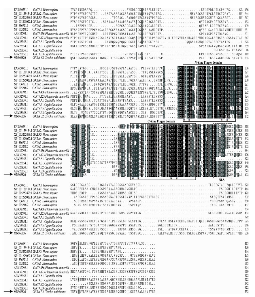
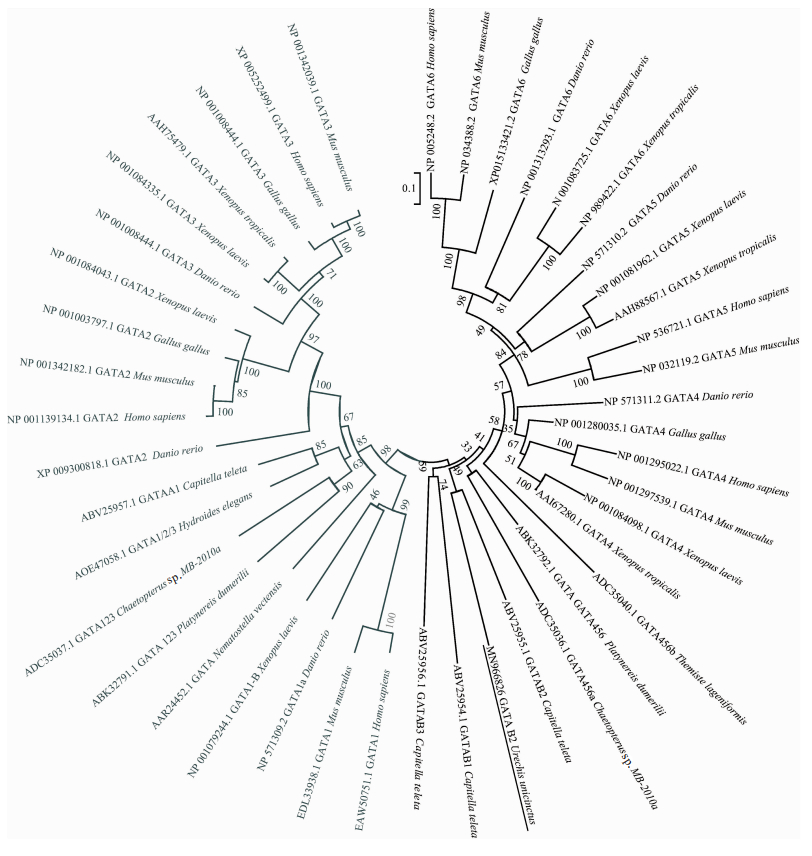
2 结果 2.1 UuGATA B2的序列特征分析UuGATA B2的开放阅读框(Open reading frame, ORF)全长为1 788 bp(GenBank accession: MN966826),编码585个氨基酸,预测的蛋白分子质量为61.54 kD,等电点为9.74。多序列比对显示UuGATA B2的氨基酸序列中具有两个锌指结构域,且每个结构域中都包含4个保守的半胱氨酸残基(见图 1)。NLS-Mapper预测该序列中具有核定位信号(Nuclear localization signal, NLS),位于其氨基酸的第449~458位,序列为QTRKRKPKSM(见图 1),SignalP和TMHMM预测显示该蛋白无跨膜结构和信号肽。进化树分析显示单环刺螠的GATA B2首先与小头虫(Capitella teleta)的GATA B2聚为一支,再与其它无脊椎动物的GATA4/5/6亚家族成员聚为一支,然后与脊椎动物的GATA4/5/6亚家族聚类,最后与GATA1/2/3亚家族聚类(见图 2)。以上结果初步确定本实验获得的序列为单环刺螠的GATA B2序列。
|
(黑色阴影表示相同的氨基酸,深灰色阴影表示氨基酸的一致性为70%以上,浅灰色阴影表示氨基酸的一致性为50%以上;黑色框标记锌指结构域;星号(*)标记半胱氨酸残基;黑色虚线框标记核定位信号。Black shaded regions are the same amino acids, the dark gray shaded regions are 70% identity amino acids, the light gray shaded regions are 50%; the zinc finger domain is boxed in black, the asterisk (*) marks the cysteine residue; the NLS is marked with a black dotted box. ) 图 1 不同物种GATA家族成员氨基酸的多序列比对 Fig. 1 Multiple sequence alignment of GATA proteins from different species |
|
(灰色为GATA1/2/3亚家族,黑色为GATA4/5/6亚家族;横线标识单环刺螠的GATA B2。Gray is the GATA1/2/3 subfamily, black is the GATA4/5/6 subfamily; horizontal line marks UuGATA B2. ) 图 2 不同物种GATA成员的系统进化分析 Fig. 2 Phylogenetic analysis of GATA family among the different species |
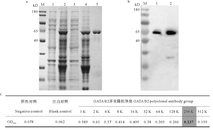
本实验成功诱导UuGATA B2重组蛋白的表达,SDS-PAGE结果显示该重组蛋白主要以包涵体的形式表达,大小约63 kD,与预测的单环刺螠GATA B2蛋白分子质量一致(见图 3a)。将诱导7 h的原核表达菌液进行超声破碎并经Ni-NTA柱亲和层析纯化获得纯度超过85%的重组蛋白(见图 3a)。
|
(a. UuGATA B2的原核表达和蛋白纯化。M:蛋白分子量标准;1:未诱导菌;2:诱导7 h菌液;3:超声后可溶蛋白;4:超声后的包涵体蛋白;5:Ni-NTA柱纯化后的蛋白。b. Western blotting检测UuGATA B2抗体的特异性。1:纯化蛋白; 2:后肠总蛋白。c.间接ELSIA实验检测UuGATA B2抗体的效价。灰色阴影框为测得的UuGATA B2抗体效价(大于阴性对照2.1倍最高的稀释倍数)。a. Prokaryotic expression and purification of UuGATA B2 protein. M: Protein molecular mass marker; 1: The product without IPTG; 2: The induced products at 7 h; 3: The supernatant of ultrasonicated product; 4: The inclusion body of ultrasonicated product; 5: Purified GATA B2 with Ni-NTA. b. The specificity of anti-UuGATA B2 was detected by Western blotting. 1: the purified protein; 2: Total proteins from hindgut. c. The titer of anti-UuGATA B2 was detected by indirect ELISA. The gray shaded box was titer of anti-UuGATA B2 which was the highest dilution ratio greater than 2.1 times of the negative control. ) 图 3 单环刺螠GATA B2蛋白的原核表达、蛋白纯化和抗体检测 Fig. 3 Prokaryotic expression and purification ofUuGATA B2 protein and detection of UuGATA B2 polyclonal antibody |
利用Western blotting检测UuGATA B2抗体的特异性,结果显示在单环刺螠后肠总蛋白和UuGATA B2纯化蛋白中均产生一个清晰的主带,其大小与目的蛋白的大小相符(见图 3b)。间接ELISA检测GATA B2的抗体效价为1:2.56×10⁵(见图 3c)。以上检测表明UuGATA B2多克隆抗体具有特异性和较高的效价,可以用于后续实验。
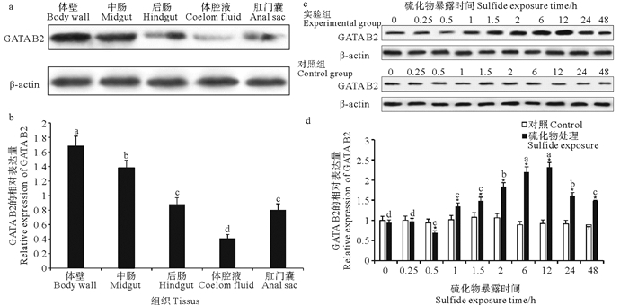
2.3 UuGATA B2的组织分布和硫化物应激下的表达分析用上述制备的UuGATA B2多克隆抗体,使用Western blotting技术检测UuGATA B2在不同组织(体壁、中肠、后肠、体腔液和肛门囊)中的表达(见图 4a、b)。结果显示该蛋白在体壁中的含量最高,中肠次之,后肠和肛门囊中表达量较低,体腔液中最低。
|
(a. Western blotting检测GATA B2在单环刺螠不同组织中的表达量; b. a图转换的灰度值,依据a图中β-actin的表达量计算UuGATA B2的相对表达量; c. UuGATA B2在硫化物暴露下的单环刺螠后肠总蛋白中的含量; d. c图转换的灰度值。β-actin为内参蛋白;所有数据均以平均值±标准误(n=3)表示。不同字母表示不同组织间或硫化物处理组间存在显著性差异(P < 0.05);表示硫化物处理组与同时刻对照组数据之间存在显著差异(P < 0.05)。a. The expression of UuGATA B2 in different tissue was detected by Western blotting; b. The converted grayscal value from figure a, the relative expression levels of UuGATA B2 are normalized to β-actin according to the result of a; c. Content of UuGATA B2 in total protein of the hindgut from U. unicinctus exposed to sulfide; d. The converted grayscale value from figure c. β-actin is the internal control protein. Data are indicated as mean ± SD (n=3). Different letters indicate significant difference among the sulfide exposure group or different tissues (P < 0.05); represents significant difference (P < 0.05) between the sulfide exposure group and control at the same time. ) 图 4 单环刺螠GATA B2在不同组织(a、b)和150 μmol/L硫化物暴露后后肠中的表达量(c、d) Fig. 4 Content of UuGATA B2 in different tissues (a, b) and the hindgut from U. unicinctus exposed to 150 μmol/L sulfide (c, d) |
单环刺螠后肠是重要的呼吸器官,直接与海水接触,是硫化物代谢的重要组织器官[27];且在150 μmol/L硫化物应激过程中,单环刺螠后肠中硫代谢关键基因sqr mRNA的表达量最高可达对照组的50倍,较体壁、中肠、肛门囊等组织应激反应更为明显[24, 28]。为探究UuGATA B2的硫化物应激特征,作者检测了其在150 μmol/L硫化物暴露条件下的后肠中的表达(见图 4c、d)。结果显示UuGATA B2在未经硫化物处理的单环刺螠后肠总蛋白中便呈现一定的含量,在硫化物暴露0.5 h时UuGATA B2的含量显著性降低,为对照组的70%;随后UuGATA B2含量呈现时间依赖特征,即随着暴露时间的延长其含量逐步升高,当硫化物暴露12 h时,UuGATA B2含量达到最高,是对照组的2.32倍;之后,UuGATA B2在后肠总蛋白中的含量有所降低,但仍显著高于对照组。实验期间,不同时间点对照组中的UuGATA B2含量未见显著性变化。
3 讨论本文通过扩增获得UuGATA B2的ORF全长为1 788 bp,编码585个氨基酸。该蛋白具有核定位信号,不具有信号肽和跨膜结构,包含两个GATA家族特异的锌指结构域(见图 1),N端的锌指结构域用于稳定与特异序列的结合或与其它蛋白相互结合,C端的锌指结构域用于识别特异序列T/A(GATA)A/G[29-30],以上特征表明其具有GATA转录因子的基本特征。系统进化分析表明其应为GATA4/5/6亚家族成员。
在单环刺螠成体各组织中,UuGATA B2在体壁的表达量最高,中肠次之,后肠和肛门囊的表达量较低,体腔液中最低(见图 4a、b)。研究表明单环刺螠的体壁含有中胚层发育的肌肉层,也是抵御环境中硫化物的第一道屏障[24, 27, 31];单环刺螠的肠道也是中内胚层发育的产物[32],说明UuGATA B2在单环刺螠成体中主要分布于中内胚层相关的组织。GATA4/5/6亚家族在脊椎动物中主要参与中内胚层相关器官的形成发育和功能维持,包括在肠道、心血管系统等[33-34]。在果蝇和线虫中,该亚家族的成员也是肠道等中内胚层相关器官发育和功能维持的关键因子[10, 35-36]。说明UuGATA B2在中内胚层的功能具有保守性。
GATA家族作为经典的转录因子家族,除了在胚层分化和相关器官的形成和功能维持中发挥重要作用外,在少数物种中还报道其参与环境的应激反应,除秀丽隐杆线虫的GATA4/5/6亚家族的ELT-2和ELT-3外[16-21],酿酒酵母(Saccharomyces cerevisiae)在盐胁迫下,GATA转录因子GLN3和GAT1的缺失导致盐诱导相关基因表达的减少[37];粟酒裂殖酵母(Schizosaccharomyces pombe)在缺氮条件下,GATA转录因子GAF1会下调性发育相关基因ste11的表达[38];构巢曲霉(Aspergillus nidulans)的碳胁迫也需要GATA转录因子的参与[39]。本文采用免疫印迹检测了其在150 μmol/L硫化物暴露的单环刺螠后肠中的UuGATA B2含量变化(见图 4c、d)。在硫化物暴露的后肠中,UuGATA B2在暴露0.5 h时显著性降低,可能是硫化物对其合成产生了影响;但在暴露1 h后,其含量呈现时间依赖性,即随着硫化物暴露时间的增加,UuGATA B2的含量逐渐升高以应对高浓度硫化物的影响。这一结果首次证明了GATA B2可以参与硫化物应激过程。将GATA B2与已鉴定的sqr转录因子(HSF1、NF1、SP1、RWDD1)对比发现,在应激起始时间上,GATA B2的起始应激时间是0.5 h,是最早响应硫化物胁迫的转录因子;在应激最高峰的时间上,GATA B2与RWDD1一致,为12 h,早于其它转录因子的24 h;在蛋白含量增加的幅度上,HSF1在24 h的增加幅度最高(对照组的7倍),GATA B2在12 h最低(对照组的2.32倍)[24-25, 40]。以上分析说明GATA B2在响应硫化物胁迫的时间和应激最高峰的时间上先于其它转录因子,而这些转录因子在硫化物暴露中的不同应激特征也暗示sqr的转录可能存在复杂的调控机制,GATA B2对sqr的转录调控以及多转录因子的协同调控机制也是本研究团队后续探究硫代谢调控的重要研究方向。
| [1] |
Merika M N, Orkin S H. DNA-binding specificity of GATA family transcription factors[J]. Molecular and Cellular Biology, 1993, 13(7): 3999-4010. DOI:10.1128/MCB.13.7.3999
(  0) 0) |
| [2] |
Patient R K, McGhee J D. The GATA family (vertebrates and invertebrates)[J]. Current Opinion in Genetics & Development, 2002, 12(4): 416-422.
( 0) |
| [3] |
Tsai S F, Martin D I, Zon L I, et al. Cloning of cDNA for the major DNA-binding protein of the erythroid lineage through expression in mammalian cells[J]. Nature, 1989, 339(6224): 446-451. DOI:10.1038/339446a0
( 0) |
| [4] |
Lowry J A, Atchley W R. Molecular evolution of the GATA family of transcription factors: Conservation within the DNA-binding domain[J]. Journal of Molecular Evolution, 2000, 50(2): 103-115. DOI:10.1007/s002399910012
( 0) |
| [5] |
Tremblay M, Sanchez-Ferras O, Bouchard M, et al. GATA transcription factors in development and disease[J]. Development, 2018, 145(20): dev164384. DOI:10.1242/dev.164384
( 0) |
| [6] |
Whitcomb J, Gharibeh L, Nemer M. From embryogenesis to adulthood: Critical role for GATA factors in heart development and function[J]. IUBMB Life, 2020, 72(1): 53-67. DOI:10.1002/iub.2163
( 0) |
| [7] |
翁健俏, 贾凡, 徐梅, 等. GATA3对耳蜗发育和功能维持的作用[J]. 杭州师范大学学报(自然科学版), 2020, 19(5): 496-502. Weng J Q, Jia F, Xu M, et al. Effects of GATA3 on cochlear development and function maintenance[J]. Journal of Hangzhou Normal University (Natural Science Edition), 2020, 19(5): 496-502. ( 0) |
| [8] |
Gillis W Q, Bowerman B A, Schneider S Q. The evolution of protostome GATA factors: Molecular phylogenetics, synteny, and intron/exon structure reveal orthologous relationships[J]. BMC Evolutionary Biology, 2008, 8(1): 1-15. DOI:10.1186/1471-2148-8-1
( 0) |
| [9] |
Klinedinst S L. The Functions of the GATA Factor Pannier and its Partner U-Shaped in Specifying the Drosophila Heart[D]. Ann Arbor: University of Michigan, 2006.
( 0) |
| [10] |
Dimov I, Maduro M F. The C. elegans intestine: Organogenesis, digestion, and physiology[J]. Cell and Tissue Research, 2019, 377: 383-396. DOI:10.1007/s00441-019-03036-4
( 0) |
| [11] |
Gillis W J, Bowerman B, Schneider S Q. Ectoderm-and endomesoderm-specific GATA transcription factors in the marine annelid Platynereis dumerilli[J]. Evolution & Development, 2007, 9(1): 39-50.
( 0) |
| [12] |
Boyle M J, Seaver E C. Developmental expression of foxA and gata genes during gut formation in the polychaete annelid, Capitella sp. Ⅰ[J]. Evolution & Developmen, 2008, 10(1): 89-105.
( 0) |
| [13] |
Jiang H, Wang D. The microbial zoo in the C. elegans intestine: Bacteria, fungi and viruses[J]. Viruses, 2018, 10(2): 85. DOI:10.3390/v10020085
( 0) |
| [14] |
Yue F, Zhou Z, Wang L, et al. A conserved zinc finger transcription factor GATA involving in the hemocyte production of scallop Chlamys farreri[J]. Fish & Shellfish Immunology, 2014, 39(2): 125-135.
( 0) |
| [15] |
Song X, Xin X, Dong M, et al. The ancient role for GATA2/3 transcription factor homolog in the hemocyte production of oyster[J]. Developmental & Comparative Immunology, 2018, 82: 55-65.
( 0) |
| [16] |
Shao H, Wang D. Long-term and low-dose exposure to nanopolystyrene induces a protective strategy to maintain functional state of intestine barrier in nematode Caenorhabditis elegans[J]. Environmental Pollution, 2020, 258: 113649. DOI:10.1016/j.envpol.2019.113649
( 0) |
| [17] |
Roh H C, Dimitrov I, Deshmukh K, et al. A modular system of DNA enhancer elements mediates tissue-specific activation of transcription by high dietary zinc in C. elegans[J]. Nucleic Acids Research, 2015, 43(2): 803-816. DOI:10.1093/nar/gku1360
( 0) |
| [18] |
Schieber M, Chandel N S. TOR signaling couples oxygen sensing to lifespan in C. elegans[J]. Cell Reports, 2014, 9(1): 9-15. DOI:10.1016/j.celrep.2014.08.075
( 0) |
| [19] |
Mesbahi H, Pho K B, Tench A J, et al. Cuticle collagen expression is regulated in response to environmental stimuli by the GATA transcription factor ELT-3 in Caenorhabditis elegans[J]. Genetics, 2020, 215(2): 483-495. DOI:10.1534/genetics.120.303125
( 0) |
| [20] |
Hu Q, D'Amora D R, MacNeil L T, et al. The oxidative stress response in Caenorhabditis elegans requires the GATA transcription factor ELT-3 and SKN-1/Nrf2[J]. Genetics, 2017, 206(4): 1909-1922. DOI:10.1534/genetics.116.198788
( 0) |
| [21] |
Arp A J, Hansen B M, Julian D. Burrow environment and coelomic fluid characteristics of the echiuran worm Urechis caupo from populations at three sites in northern California[J]. Marine Biology, 1992, 113(4): 613-623. DOI:10.1007/BF00349705
( 0) |
| [22] |
李诺, 宋淑莲, 唐永政. 单环刺螠生活史研究[J]. 齐鲁渔业, 1995(6): 24-27. Li N, Song S L, Tang Y Z. The life history of Urechis unicinctus[J]. Shandong Fisheries, 1995(6): 24-27. ( 0) |
| [23] |
张志峰, 王思锋, 霍继革, 等. 单环刺螠对硫化物暴露的呼吸代谢适应[J]. 中国海洋大学学报(自然科学版), 2006, 36(4): 639-644. Zhang Z F, Wang S F, Huo J G, et al. Adaptation of respiratory metabolism to sulfide exposure in Urechis unicinctus[J]. Periodical of Ocean University of China (Natural Science), 2006, 36(4): 639-644. ( 0) |
| [24] |
Ma Y B, Zhang Z F, Shao M Y, et al. Response of sulfide quinone oxidoreductase to sulfide exposure in the echiuran worm Urechis unicinctus[J]. Marine Biotechnology, 2012, 14(2): 245-251. DOI:10.1007/s10126-011-9408-1
( 0) |
| [25] |
Li X, Liu X, Qin Z, et al. A novel transcription factor Rwdd1 and its SUMOylation inhibit the expression of sqr, a key gene of mitochondrial sulfide metabolism in Urechis unicinctus[J]. Aquatic Toxicology, 2018, 204: 180-189. DOI:10.1016/j.aquatox.2018.09.012
( 0) |
| [26] |
Liu X, Zhang Z, Ma X, et al. Sulfide exposure results in enhanced sqr transcription through upregulating the expression and activation of HSF1 in echiuran worm Urechis unicinctus[J]. Aquatic Toxicology, 2016, 170: 229-239. DOI:10.1016/j.aquatox.2015.11.021
( 0) |
| [27] |
Arp A J, Menon J G, Julian D. Multiple mechanisms provide tolerance to environmental sulfide in Urechis caupo[J]. American Zoologist, 1995, 35(2): 132-144. DOI:10.1093/icb/35.2.132
( 0) |
| [28] |
Ma Y B, Zhang Z F, Shao M Y, et al. Function of the anal sacs and mid-gut in mitochondrial sulphide metabolism in the echiuran worm Urechis unicinctus[J]. Marine Biology Research, 2012, 8(10): 1026-1031. DOI:10.1080/17451000.2012.707320
( 0) |
| [29] |
Lowry J A, Atchley W R. Molecular evolution of the GATA family of transcription factors: Conservation within the DNA-binding domain[J]. Journal of Molecular Evolution, 2000, 50(2): 103-115. DOI:10.1007/s002399910012
( 0) |
| [30] |
He C, Cheng H, Zhou R. GATA family of transcription factors of vertebrates: Phylogenetics and chromosomal synteny[J]. Journal of Biosciences, 2007, 32(3): 1273-1280.
( 0) |
| [31] |
李诺, 宋淑莲, 唐永政. 单环刺螠[J]. 生物学通报, 1998(8): 3-5. Li N, Song S L, Tang Y Z. Urechis unicinctus[J]. Bulletin of Biology, 1998(8): 3-5. ( 0) |
| [32] |
陈宗涛, 张志峰, 康庆浩, 等. 单环刺螠消化道的发生和分化[J]. 中国水产科学, 2006(5): 700-707. Chen Z T, Zhang Z F, Kang Q H, et al. Journal of Fishery Sciences of China, 2006(5): 700-707[J]. Journal of Fishery Sciences of China, 2006(5): 700-707. DOI:10.3321/j.issn:1005-8737.2006.05.002 ( 0) |
| [33] |
Beuling E, Baffour-Awuah N Y A, Stapleton K A, et al. GATA factors regulate proliferation, differentiation, and gene expression in small intestine of mature mice[J]. Gastroenterology, 2011, 140(4): 1219-1229. DOI:10.1053/j.gastro.2011.01.033
( 0) |
| [34] |
Walker E M, Thompson C A, Battle M A. GATA4 and GATA6 regulate intestinal epithelial cytodifferentiation during development[J]. Developmental Biology, 2014, 392(2): 283-294. DOI:10.1016/j.ydbio.2014.05.017
( 0) |
| [35] |
Murakami R, Okumura T, Uchiyama H. GATA factors as key regulatory molecules in the development of Drosophila endoderm[J]. Development, Growth & Differentiation, 2005, 47(9): 581-589.
( 0) |
| [36] |
Martínez-Corrales G, Cabrero P, Dow J A T, et al. Novel roles for GATAe in growth, maintenance and proliferation of cell populations in the Drosophila renal tubule[J]. Development, 2019, 146: dev178087. DOI:10.1242/dev.178087
( 0) |
| [37] |
Crespo J L, Daicho K, Ushimaru T, et al. The GATA transcription factors GLN3 and GAT1 link TOR to salt stress in Saccharomyces cerevisiae[J]. Journal of Biological Chemistry, 2001, 276(37): 34441-34444. DOI:10.1074/jbc.M103601200
( 0) |
| [38] |
Kim L, Hoe K L, Yu Y M, et al. The fission yeast GATA factor, Gaf1, modulates sexual development via direct down-regulation of ste11+ expression in response to nitrogen starvation[J]. PloS One, 2012, 7(8): e42409. DOI:10.1371/journal.pone.0042409
( 0) |
| [39] |
Macios M, Caddick M X, Weglenski P, et al. The GATA factors AREA and AREB together with the co-repressor NMRA, negatively regulate arginine catabolism in Aspergillus nidulans in response to nitrogen and carbon source[J]. Fungal Genetics and Biology, 2012, 49(3): 189-198. DOI:10.1016/j.fgb.2012.01.004
( 0) |
| [40] |
Liu X, Qin Z, Li X, et al. NF1, Sp1 and HSF1 are synergistically involved in sulfide-induced sqr activation in echiuran worm Urechis unicinctus[J]. Aquatic Toxicology, 2016, 175: 232-240. DOI:10.1016/j.aquatox.2016.04.002
( 0) |